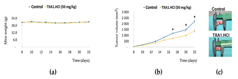
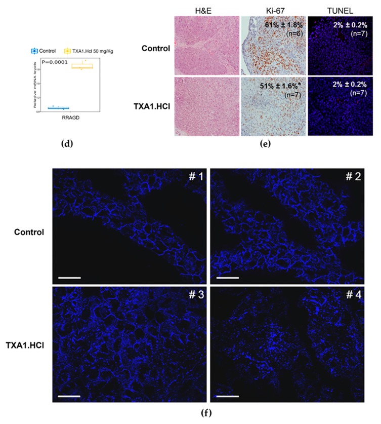
Figure 9.
TXA1.HCl decreases tumor growth and proliferation and affects cholesterol localization and RAGD expression in NCI-H460 tumor xenografts in nude mice. Nude mice bearing tumor xenografts (40–80 mm3) were s.c. injected three times per week with TXA1.HCl (50 mg/kg, n = 7) or vehicle control (saline, n = 7). (a) Body weight changes in mice during treatment. Results are expressed as the mean ± SEM, n = 7. * p < 0.05 vs. control. (b) Tumor growth curves. Tumor dimensions were measured using calipers and volumes inferred on the indicated days as previously mentioned. (c) Representative tumors resected at the end of the experiment (at day 22 after cells inoculation). Caliper is present to compare the tumor sizes. Since some tumors were bilobulated, each lobule was measured alone and the total volume was inferred. (d) Tumors histological analysis. Representative images of tumor xenografts sections: H&E stain, immunohistochemistry for Ki-67 and TUNEL fluorescence microscopy. Original magnification is 40x. (e) RagD expression levels, analyzed by quantitative real-time PCR. Values are expressed after normalization for an endogenous control (Hprt1). Data plotted with ggplot and p values were calculated using t. test in R statistical programming language, comparing TXA1.Hcl treatment vs control. Results are expressed as mean ± SEM, for n = 3. * p < 0.05 vs. control. (f) Cholesterol localization was evaluated by filipin staining. Images are representative of tumor xenografts from the different animals (control mice #1 and #2; treated mice #3 and #4). Bar = 20 µM.


