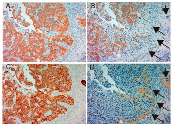
Figure 3.

Immunohistochemical appearance of total MET (A), phospho-MET (B), matriptase (C) and HAI-1 (D) in serial tissue sections of high-grade urothelial carcinoma (patient 23, ×200 magnification). Dominant expression of total MET (A) and matriptase (C) is observed. Phosphorylation of MET is also upregulated (B); however, downregulation is observed in part (arrows). In contrast, increased expression of HAI-1 is observed in the area with downregulated phospho-MET (D, allows). This case shows a representative pattern of reciprocal expression of HAI-1 and phospho-MET.
