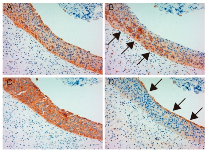
Figure 4.

Immunohistochemical appearance of total MET (A), phospho-MET (B), matriptase (C) and HAI-1 (D) in serial tissue sections of normal urothelium (patient 23, ×200 magnification). Total MET and matriptase are expressed in the majority of normal urothelial cells (A,C). However, phosphorylation of MET is upregulated in the basal area of the urothelium (B, arrows), and HAI-1 is expressed in umbrella cells (D). These patterns are representative of non-malignant urothelium adjacent to a cancer lesion.
