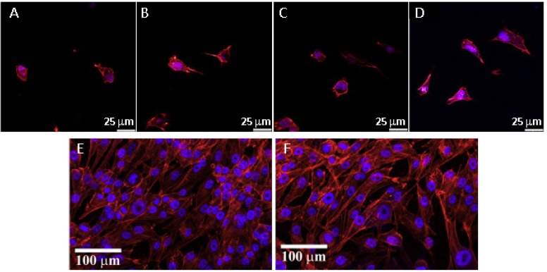
Figure 2.
Fluorescence microscope images of adipose-derived mesenchymal stem cells cultured for 6 h on HAp with variable surface topography; control (A), nanosheet (B), nanorod (C) and micro–nanohybrid (D). MG63 human osteoblasts cultured for 15 days on zirconia (E) or alumina–zirconia particulate composites (F). Actin filaments are stained in red, while the cell nuclei are stained in blue. Adapted from [30,35] with permission from Elsevier.

