Figure 2.
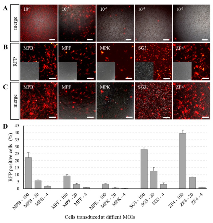
Cells transduced with recombinant baculovirus. (A) Titer determination by end-point dilution method based on RFP fluorescence. Baculoviral stocks diluted from 10−2 to 10−8 times with 10 μL were added to a 96-well plate. The number on the upper left corner of each image is the volume (μL) of original baculoviral stock added to each well. Scale bars, 400 μm. (B,C) Expression of RFP in Mylopharyngodon piceus bladder (MPB), fin (MPF), and kidney (MPK); Oryzias latipes spermatogonia (SG3); and Danio rerio embryonic fibroblast (ZF4) cells transduced with BV-CMV-ie1-pr at a multiplicity of infection (MOI) of 20. The expression of RFP was observed under an inverted fluorescence microscope after 3 days of transduction. Scale bars, 200 μm. (D) Transduction efficiencies of BV-CMV-ie1-pr in MPB, MPF, MPK, SG3, and ZF4 cells at different MOIs. The transduced efficiencies were determined by counting RFP-positive cells under an inverted fluorescence microscope at 3 days post-transduction in triplicate. The number after the cell name is the corresponding incubation dose in MOI. Values are indicated as mean ± SD.

