Figure 2.
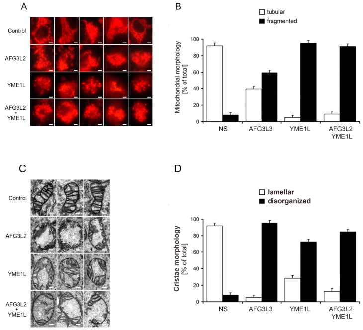
Loss of AFG3L2 and/or YME1L leads to mitochondrial fragmentation and severely disorganized and attenuated cristae architecture. (A) AFG3L2, YME1L, and AFG3L2/YME1L knockdown cells exhibit markedly fragmented mitochondrial reticulum. Cells were analyzed using a Nikon Diaphot 200 inverted microscope equipped with an Olympus DP50 camera. Bar, 10 μm. (B) The quantification of mitochondrial network morphology in control and KD cells. Cells containing tubular (white bars) or fragmented (black bars) mitochondria were counted in a double-blind manner. More than 100 cells were scored per experiment. (C) AFG3L2, YME1L, and AFG3L2/YME1L KD cells exhibit markedly altered and attenuated cristae architecture. The cells were incubated in PBS containing 2% potassium permanganate for 15 min, washed with PBS, and dehydrated with an ethanol series. They were then embedded in Durcupan Epon, sectioned by microtome to thicknesses ranging from 600 to 900 Å, and stained with lead citrate and uranyl acetate. The sections were viewed with a JEOL JEM-1200 EX transmission electron microscope. Bars correspond to 200 nm. (D) The quantification of cristae morphology in KD cells. Approximately 50 sections of individual cells were scored in a double-blind manner. Error bars correspond to SD from the mean.

