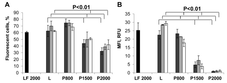
Figure 3.
Liposomes-mediated delivery of plasmid DNA in HEK 293 cells measured by EGFP expression. The percentage of EGFP-positive cells (A) and mean fluorescence intensity (MFI) of the cell population (B) were evaluated by flow cytometry 48 h after transfection. Cells were incubated with the lipoplexes formed by liposomes and pEGFP-C2 (2 μg/mL) at N/P ratios of 4/1 (dark grey bars), 6/1 (light grey bars) or 8/1 (white bars) in the presence of 10% FBS. LF2000- Lipofectamine 2000 (black bars) was used under optimal conditions. The differences between mean values for L and other preparations at the same N/P with p < 0.01 were considered statistically significant according to Mann-Whitney U-test.

