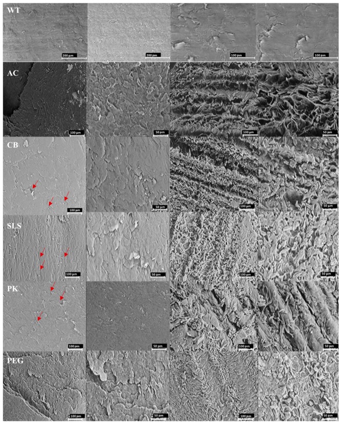
Figure 1.
Scanning electron microscopy photomicrographs at different magnifications of the healthy nail plates incubated with the enhancers (WT: nail without treatment, AC: acetylcysteine, CB: carbocysteine, SLS: sodium lauryl sulfate, PK: potassium phosphate, and PEG: polyethylene glycol 300) on their external (dorsal) surface (two first left columns) and on the internal (ventral) surface (two right columns). Red arrows point to some of the pores.

