Figure 6.
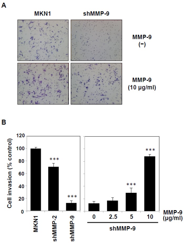
Potentiation of the invasion of MMP-9-knockdown MKN1 cells by exogenous MMP-9. MMP-2- or MMP-9-knockdown MKN1 cells were prepared by RNA interference as described in the Materials and Methods section. (A) The MMP-9-knockdown MKN1 cells (right) and parent cells (left) were assayed for in vitro invasion using Matrigel basement membranes in the presence or absence of MMP-9 purified from THP-1 cells. After culturing for 16 h, the cells that had migrated into the lower chamber were microscopically observed (40×). (B) MMP-2- or MMP-9-knockdown MKN1 cells were subjected to an invasion assay, and the migrated cells were counted under a microscope (Left). The invasion assay was performed in the presence of purified MMP-9 (2.5–10 μg/mL) (right). Experiments were performed in triplicate, and the data are presented as the mean ± SEM. Statistical data analysis was conducted using the Student’s t-test. *** p < 0.005.

