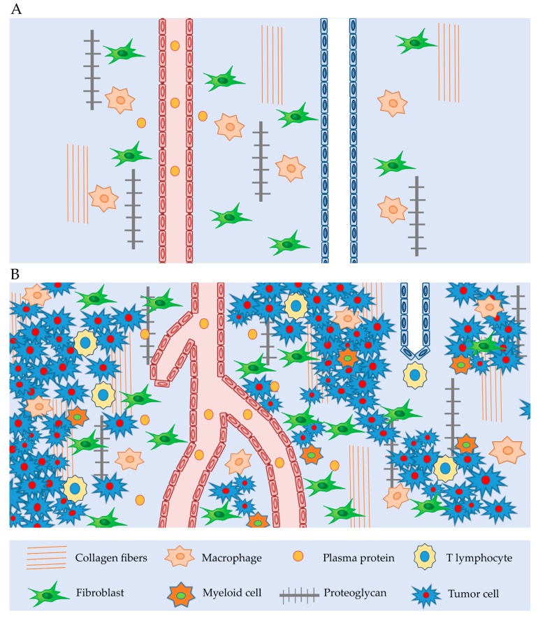
Figure 1.
Interstitium tissue: (A) Normal interstitium tissue with lymphatic and blood vessel, macrophages, fibroblast, collagen fibers, proteoglygan. (B) Tumor interstitium tissue with reduced lymphatic vessel, winding blood vessel, macrophages, fibroblasts, increased amount of extravasated plasma proteins, T-lymphocyte, myeloid cells, collagen fibers, proteoglygan and tumor cells, all of which induce to an increase in tumor interstitial pressure.

