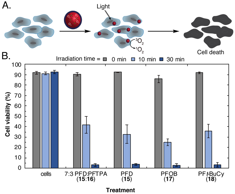
Figure 3.
(A) Photodynamic therapy with perfluorocarbon nanoemulsions containing 13 (0.5 mM). Cells were incubated with PFC emulsions containing 13, washed via centrifugation, and irradiated (420 nm, 8.5 mW/cm2) for 0 min (grey), 10 min (light blue), or 30 min (dark blue). (B) Flow cytometry analysis after light treatment. After incubation, washing, and light treatment, cells were stained with propidium iodide and analyzed by flow cytometry to determine the degree of cell death. Dead cells were characterized as exhibiting fluorescence >102 (Figure S4). Error bars represent the standard deviation of 3 replicate samples.

