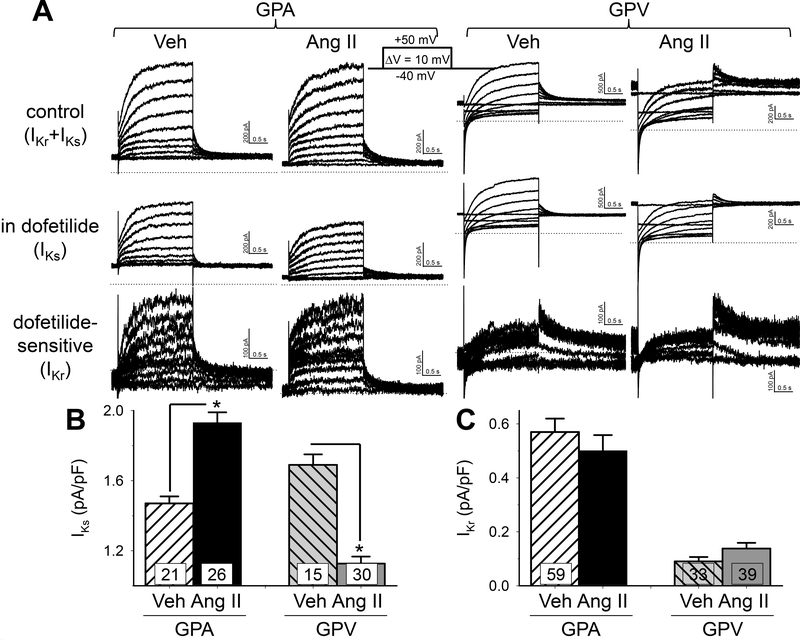
Fig. 2. Effects of chronic Ang II administration on the slow and rapid delayed rectifier currents (IKs and IKr) of guinea pig atrial and ventricular myocytes.
(A) Representative current traces recorded from GPA and GPV myocytes from Veh and Ang II animals. Currents were elicited by the diagrammed voltage clamp protocol. Top row: control (Ικγ+Iks)· Second row: in 1 uM dofetilide (IKs)· Subtracting IKs from control produced dofetilide-sensitive current (third row, ΙΚγ). (B) and (C) Summary of lKs and lKr current density in GPA and GPV myocytes from Veh and Ang ll animals. For each cell, lKs was measured as peak tail current amplitude at −20 mV following a 2-s pulse to +40 mV, and lKr was measured as peak tail current amplitude at −60 mV following a 2-s pulse to +20 mV. Validation of such measurements is presented in Fig. S1. Both were normalized by cell capacitance. * p < 0.05 Ang ll vs Veh.

