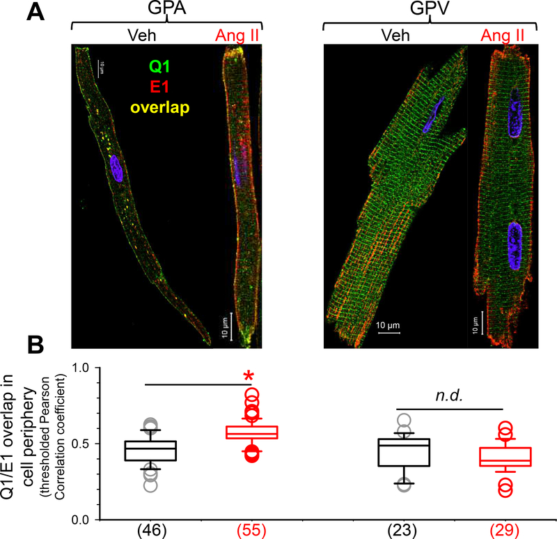
Fig. 4. Effects of chronic Ang II administration on subcellular distribution of KCNQ1 and KCNE1 in guinea pig atrial and ventricular myocytes.
(A) Confocal images of native KCNQ1 and KCNE1 detected as Alexa488 ‘green’ and Alexa568 ‘red’, respectively. (B) Box plot of degree of KCNQ1/KCNE1 overlap in cell periphery, quantified by Pearson correlation coefficients of immunofluorescence signals that were above threshold (pixels in cell free areas). Numbers of myocytes analyzed are shown in parentheses; * t-test, Ang ll vs Veh, p < 0.05.

