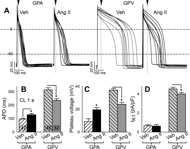
Fig. 5. Effects of chronic Ang II administration on action potential duration, plateau voltage, and outward IK1 current densities of guinea pig atrial and ventricular myocytes.
(A) Superimposed action potential traces (APs) recorded from 8–10 each of 4 groups of myocytes. Cycle length (CL) was 1 s. Dashed horizontal lines denote 0 and −60 mV. Arrowheads and dotted vertical lines denote the time point when plateau voltages were measured. (B) Action potential duration (APD) measured as interval between upstroke and when membrane voltage repolarized to −60 mV. (C) Plateau voltage measured as voltage 20 ms (GPA) or 50 ms (GPV) after the upstroke. (D) IK1 current density based on the highest value of outward IK1 current (typically at −50 mV), normalized by cell capacitance. Numbers of myocytes studied are listed (from 7 each of Veh and Ang II animals). * t-test, Ang II vs Veh, p < 0.05.

