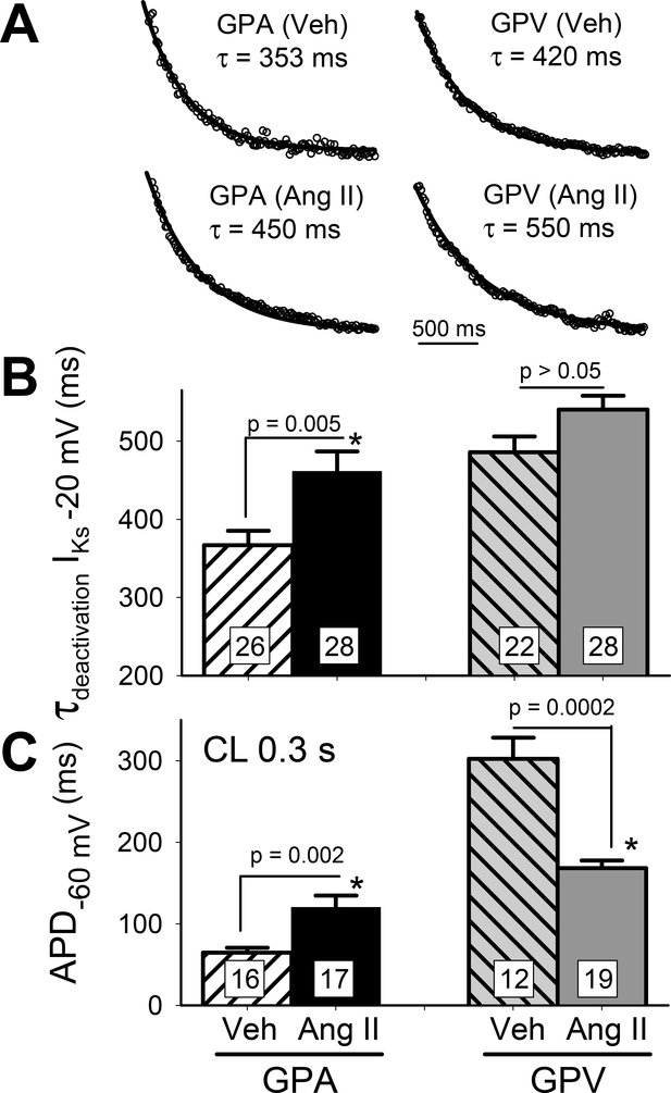
Fig. 6. chronic Ang II administration slows IKs deactivation in GPA but not GPV myocytes and this kinetic effect does not explain Ang II-induced changes in APD.
(A) Representative cases of single exponential fit (curves) to IKs tail currents at −20 mV (data points shown as open circles). Best-fit values of time constant (t) are noted. (B) Summary of τ of IKs deactivation. (C) Summary of APD at CL 0.3 s. Numbers of myocytes (from 4 animals) patch clamped are noted in n histogram bars, with p-values for t-test between Veh and Ang II noted.

