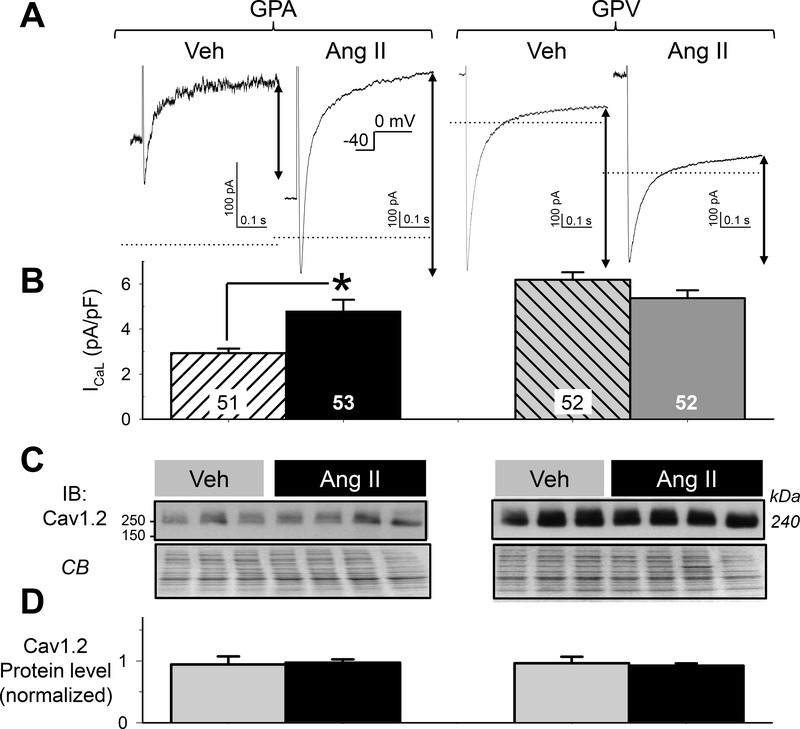
Fig. 7. Effects of chronic Ang II administration on the L-type Ca (ICaL) current densities in guinea pig atrial and ventricular myocytes.
(A) Representative current traces 4 groups of myocytes elicited by depolarizing pulses from Vh −40 mV to 0 mV. Dotted horizontal lines denote the zero current level. ICaL was quantified as the difference between the peak inward-going current and current level 500 ms into the pulse (double-arrow lines). (B) Summary of ICaL current densities. Numbers of myocytes studied are listed (from 7 each of Veh and Ang II animals). * t-test, Ang II vs Veh, p < 0.05. (C) Cav1.2 immunoblot images of whole tissue lysates (50 ug/lane) prepared from atria and left ventricles of 3 Veh and 4 Ang II animals, with CB staining as loading control. (D) Summary of densitometry. Cav1.2 band intensities were normalized by mean value of Veh lanes from the same membrane. There was no difference in Cav1.2 protein level among the 4 groups of samples.

