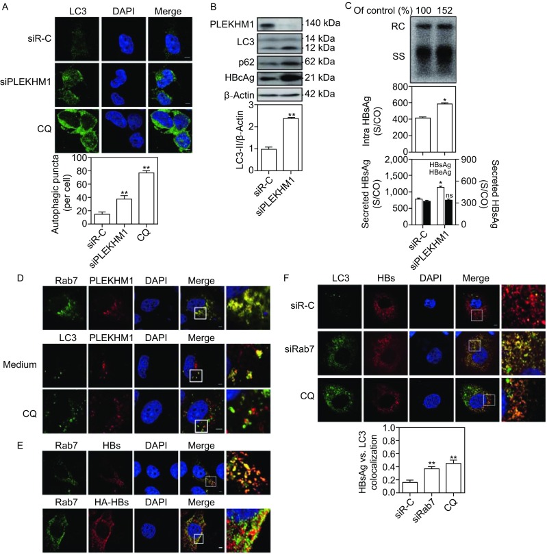
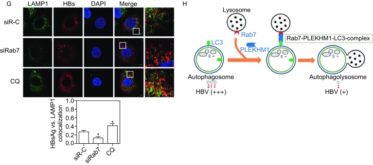
Figure 2.


Rab7 silencing interferes with the autophagic degradation of HBV by blocking the autophagosome-lysosome fusion. (A and B) HepG2.2.15 cells were transfected with 20 nmol/L siRNAs against PLEKHM1 (siPLEKHM1) or control siRNA (siR-C). After 48 h, the transfected cells were imaged by confocal microscopy. PLEKHM1, LC3, p62 and HBcAg expression were analyzed by Western blot. (C) Huh7 cells were cotransfected with the pSM2 plasmid and siRab7 or siR-C at 20 nmol/L and harvested after 72 h. Analysis of secreted HBsAg and HBeAg in culture supernatants and intracellular HBsAg from cell lysates was performed by a chemiluminescent microparticle immunoassay (CMIA). Analyses of HBV genomes in culture supernatants and HBV replicative intermediates inside the cells were separately performed as described above. (D) Huh7 cells were cotransfected with plasmids GFP-Rab7 and DesRed-PLEKHM1, or GFP-LC3 and DesRed-PLEKHM1, and harvested after 48 h. The colocalization of LC3, Rab7 and PLEKHM1 was determined by confocal microscopy. (E) Huh7 cells were firstly transfected with mCherry-HBs. After 48 h, the cells were incubated with primary antibody rabbit anti-Rab7 and then stained with Alexa Fluor 488-conjugated anti-rabbit secondary antibody IgG (upper panel). Huh7 cells were cotransfected with HA-HBs, followed by incubating with primary antibody mouse anti-HA and rabbit anti-Rab7 and then staining with Alexa Fluor 488-conjugated anti-rabbit secondary antibody IgG and Alexa Fluor 594-conjugated anti-mouse secondary antibody IgG (bottom panel). The colocalization of Rab7 and HBsAg was determined by confocal microscopy. (F and G) Huh7 cells were transfected with mCherry-HBs and harvested after 48 h. Next, the cells were fixed, incubated with primary antibody rabbit anti-LC3 or anti-LAMP1, followed by staining with Alexa Fluor 488-conjugated anti-rabbit secondary antibody IgG. Cells cultured with 10 µmol/L CQ for 48 h were used as a positive control. The colocalization of HBsAg and LC3 or LAMP1 was determined by confocal microscopy. (H) A proposed model depicting that the autophagic degradation of HBV is regulated by Rab7-PLEKHM1-LC3 complex. A part of HBsAg and HBV virions may be degraded following the fusion of autophagosomes and lysosomes. Rab7 has different roles in the transport of autophagosomes and late endosomes (LEs)/MVBs, particularly controlling the fusion process of autophagosomes with lysosomes. Silencing Rab7 and its related components led to accumulation of autophagosomes/MVBs and an increase in intracellular and released HBsAg and HBV virions due to decreased fusion to lysosomes. S/CO: signal to cutoff ratio; RC: relaxed circular DNA; SS: single-stranded DNA. The data are shown as mean ± SEM. *P < 0.05; **P < 0.01; ns, not significant
