Figure 1.

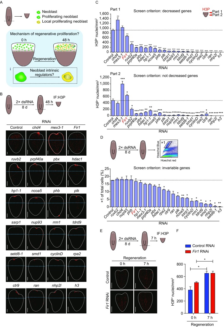
Fir1 is required for local neoblast mitosis. (A) The scientific question needed to be addressed. The mitosis adjacent to the wounds is specifically induced by tissue-missing injury (Wenemoser and Reddien, 2010), while neoblast intrinsic regulators involved in this process are still unknown. (B) Representative confocal projections through tail pieces fixed 48 h post-amputation following RNAi administration, stained with H3P antibody. The wound surfaces of tail pieces are up. Dotted lines (white): tail piece boundary. Dotted lines (blue) separate the tail pieces into two parts for quantification in (C). Unless otherwise noted, animals were fed 2× dsRNA and amputated as indicated in the cartoon (dotted red lines). Scale bars, 100 μm. (C) Mitotic density in part 1 and part 2 as separated in (B). Only one gene, Fir1, satisfies our screen criterions. In Fir1(RNAi) tail pieces mitotic density in part 1 reduced significantly, while in part 2 it was not deceased. Error bars represent SEM; * equals P < 0.05; *** equals P < 0.0001; significance determined with Student’s t test. (D) Fir1 RNAi did not affect neoblast number (percentage of X1 cells) as assayed by flow cytometry. Error bars represent SEM; * equals P < 0.05; ** equals P < 0.001; significance determined with Student’s t test. (E) Representative confocal projections through tail pieces fixed 0 h and 7 h post-amputation following RNAi knockdown of Fir1, stained with H3P antibody. Dotted lines: tail piece boundary. (F) Quantification of H3P staining in (E). These results indicate that Fir1(RNAi) do not affect the general response of neoblasts to amputation. Error bars represent SEM; * equals P < 0.05; significance determined with Student’s t test
