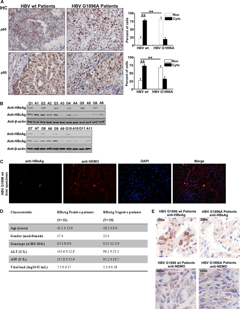
FIG 7.
Enhanced NF-κB activity in bioptic liver tissues from HBV-G1896A-infected patients. (A) Immunohistochemical staining of p65 and p50 in HBV wt- or G1896A-infected liver tumor tissues. (B) Eleven pairs of corresponding tissues were lysed and immunoblotted with the indicated antibodies. (C) Immunofluorescence in bioptic liver tissues from HBV wt-infected patients was assessed to determine the colocalization of HBeAg and NEMO. (D) Baseline characteristics of HBV-infected patients whose results are shown in panels A and C. Abbreviations: ALT, alanine aminotransferase; AST, aspartate aminotransferase. (E) Immunohistochemical staining of HBeAg or NEMO in HBV wt- or G1896A-infected liver tissues. (F) HBV templates were extracted and then amplified from the liver tissues of the HBV patients. The graphs show the sequencing results for HBV precore regions. (G) Schematic illustration of events underlying HBeAg-regulated NF-κB activity.


