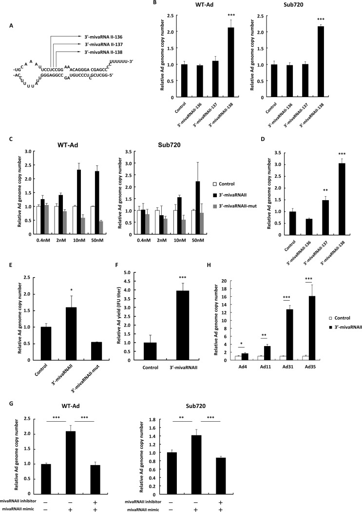
FIG 2.
3′-mivaRNAII promotes Ad replication. (A) A schematic diagram of the processing of VA-RNA II by dicer, producing 3′-mivaRNAII-136, -137, and -138. (B) HeLa cells were transfected with 3′-mivaRNAII-136, -137, or -138 mimics at 50 nM for 48 h, followed by infection with Ad (WT-Ad and Sub720) at 100 VPs/cell. Ad genome copy numbers were determined 24 h after infection and expressed as relative values (control = 1). (C) HeLa cells were transfected with 3′-mivaRNAII (3′-mivaRNAII-138) and 3′-mivaRNAII-mut mimics at the indicated concentrations for 48 h, followed by infection with Ad (WT-Ad and Sub720) at 100 VPs/cell. Ad genome copy numbers were determined 24 h after infection and expressed as relative values (control = 1 at the respective concentration). (D) A549 cells were transfected with 3′-mivaRNAII-136, -137, or -138 mimics at 50 nM for 48 h, followed by infection with WT-Ad at 100 VPs/cell. Ad genome copy numbers were determined 24 h after infection and expressed as relative values (control = 1). (E) A549 cells were transfected with 3′-mivaRNAII-138 and -mut mimics at 50 nM for 48 h, followed by infection with WT-Ad at 100 VPs/cell. Ad genome copy numbers were determined 24 h after infection and expressed as relative values (control = 1). (F) HeLa cells were transfected with 3′-mivaRNAII (3′-mivaRNAII-138) mimic at 50 nM and incubated for 48 h, followed by infection with WT-Ad at 100 VPs/cell. After 24 h of incubation, IFU titers of progeny WT-Ad in the cells were determined and expressed as relative values (control = 1). (G) HeLa cells were cotransfected with 3′-mivaRNAII (3′-mivaRNAII-138) mimic and negative control inhibitor or 3′-mivaRNAII inhibitor at 30 nM each for 48 h, followed by infection with WT-Ad at 100 VPs/cell. Ad genome copy numbers were determined 24 h after infection and expressed as relative values (control = 1). (H) HeLa cells were transfected with 3′-mivaRNAII (3′-mivaRNAII-138) mimic at 50 nM for 48 h, followed by infection with Ad4, Ad11, Ad31, or Ad35 at 100 VPs/cell. Ad genome copy numbers were determined 24 h after infection and expressed as relative values (control = 1). These data are expressed as means ± SD (n = 4).

