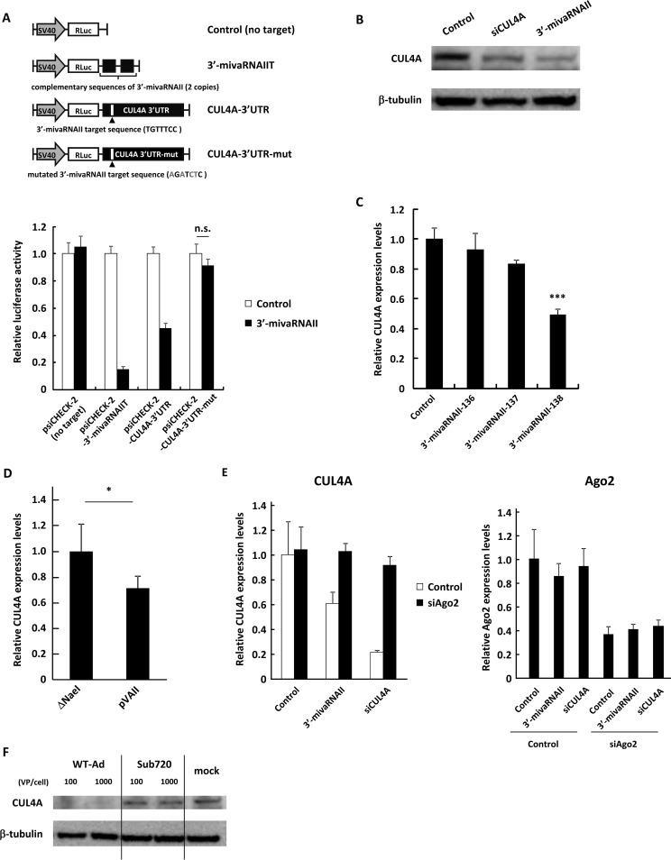
FIG 4.
3′-mivaRNAII suppresses CUL4A expression via posttranscriptional gene silencing. (A) HeLa cells were cotransfected with 3′-mivaRNAII (3′-mivaRNAII-138) mimic and the indicated reporter plasmids, described in the upper portion. Luciferase activities were determined 48 h after transfection. RLuc activities were normalized to Fluc activities. The mutated nucleotides in the sequences complementary to the seed sequences of 3′-mivaRNAII are shown in gray. SV40, SV40 promoter; n.s., not significant. (B) HeLa cells were transfected with 3′-mivaRNAII (3′-mivaRNAII-138) mimic, or siCUL4A at 50 nM. After 48 h of incubation, protein levels of CUL4A were evaluated by Western blotting analysis. (C) HeLa cells were transfected with 3′-mivaRNAII-136, -137, or -138 mimics at 50 nM. After 48 h of incubation, mRNA levels of CUL4A were determined by qRT-PCR analysis. (D) HeLa cells were transfected with ΔNaeI or pVAII. After 48 h of incubation, the mRNA levels of CUL4A were determined by qRT-PCR analysis. (E) HeLa cells were cotransfected with a 3′-mivaRNAII mimic (3′-mivaRNAII-138), siCUL4A, and siAgo2 at 50 nM. After 48 h of incubation, the mRNA levels of CUL4A and Ago2 were determined by qRT-PCR analysis. (F) HeLa cells were infected with Ad (WT-Ad and Sub720) at 100 or 1,000 VPs/cell. After 48 h of incubation, protein levels of CUL4A were evaluated by Western blotting analysis. These data are expressed as means ± SD (A and C, n = 4).

