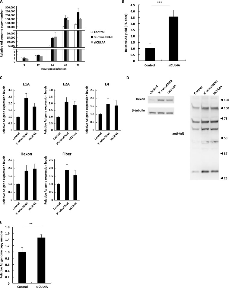
FIG 5.
Suppression of CUL4A gene expression promotes Ad infection. (A) HeLa cells were transfected with a 3′-mivaRNAII mimic (3′-mivaRNAII-138) or siCUL4A at 50 nM for 48 h, followed by infection with WT-Ad at 100 VPs/cell. After 3, 12, 24, 48, or 72 h of incubation, the Ad genome copy numbers were determined and expressed as relative values (control, 3 h postinfection = 1). (B) HeLa cells were transfected with siCUL4A at 50 nM for 48 h, followed by infection with WT-Ad at 100 VPs/cell. After 24 h of incubation, IFU titers of progeny WT-Ad were determined and expressed as relative values (control = 1). (C) HeLa cells were transfected with a 3′-mivaRNAII mimic (3′-mivaRNAII-138) or siCUL4A at 50 nM for 48 h, followed by infection with WT-Ad at 100 VPs/cell. mRNA levels of the Ad genes were determined by qRT-PCR analysis at 12 h (E1A, E2A, and E4) or 24 h (hexon and fiber) after infection. (D) HeLa cells were transfected with a 3′-mivaRNAII mimic (3′-mivaRNAII-138) or siCUL4A at 50 nM for 48 h, followed by infection with WT-Ad at 100 VPs/cell. After 24 h of incubation, the protein levels of the Ad genes were evaluated by Western blotting analysis. Numbers at the right of the membranes indicate the protein sizes in kilodaltons. Note that the sizes of the Ad major capsid proteins, hexon, penton base, and fiber, are 108 kDa, 63 kDa, and 61 kDa, respectively. (E) A549 cells were transfected with siCUL4A at 50 nM for 48h, followed by infection with WT-Ad at 100 VPs/cell. After 24 h of incubation, Ad genome copy numbers were determined and expressed as relative values (control = 1). These data are expressed as means ± SD (n = 4).

