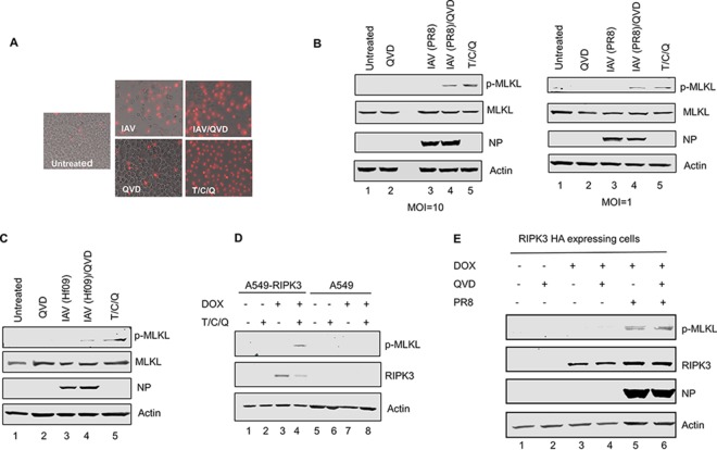
FIG 1.
IAV infection induces necroptosis in THP1 cells and the A549 cell line inducibly expressing human RIPK3. (A) Representative images of differentiated THP1 cells that were either left untreated, infected with IAV (PR8) at an MOI of 1, treated with QVD, treated with QVD for 1 h and then infected with IAV, or treated with the combination of T/C/Q. Cell death was assessed by PI staining at 16 h p.i. (B) THP1 cells were infected with IAV (PR8) at the indicated MOIs or treated as described above for panel A, and at 8 h posttreatment, cells were harvested and examined for phosphorylated MLKL by Western blotting. (C) THP1 cells were infected with IAV (Hf09) at an MOI of 10 or treated as described above for panel A. At 8 h posttreatment, cells were harvested and examined for phosphorylated MLKL by Western blotting. (D) A549 cells and an A549 cell line inducibly expressing HA-tagged human RIPK3 were cultured in the absence or presence of 0.5 μg/ml of doxycycline (DOX) for 12 h and either left untreated or treated with the combination of T/C/Q (TNF-α, cycloheximide, and QVD). At 8 h posttreatment, cells were harvested and examined for RIPK3 and phosphorylated MLKL by Western blotting. (E) The A549 cell line inducibly expressing human RIPK3 was cultured in the absence or presence of 0.5 μg/ml of doxycycline for 12 h, and cells were either left untreated or treated with QVD for 1 h and then either mock infected or infected with IAV (PR8) at an MOI of 10. At 8 h p.i., cells were harvested and examined for phosphorylated MLKL by Western blotting.

