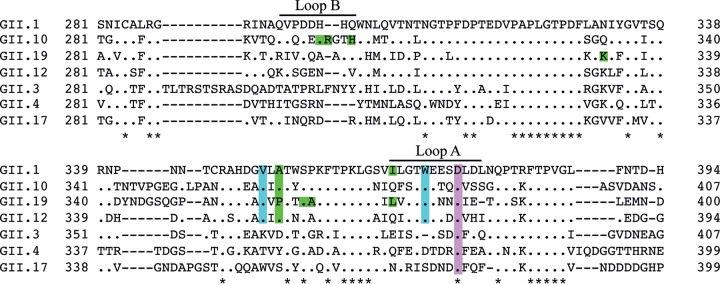
FIG 7.
Conservation of the GII bile acid binding pocket. Amino acid sequence alignments of GII capsids were performed using ClustalX (Genetyx software). The conserved P domain residues interacting with bile acid are highlighted in cyan. Variable residues that interacted with the bile acid tail are colored green. The conserved Asp residue (purple) is known to bind to the fucose moiety of HBGAs. Note that only a partial capsid sequence is shown, and the asterisks indicate highly conserved residues.

