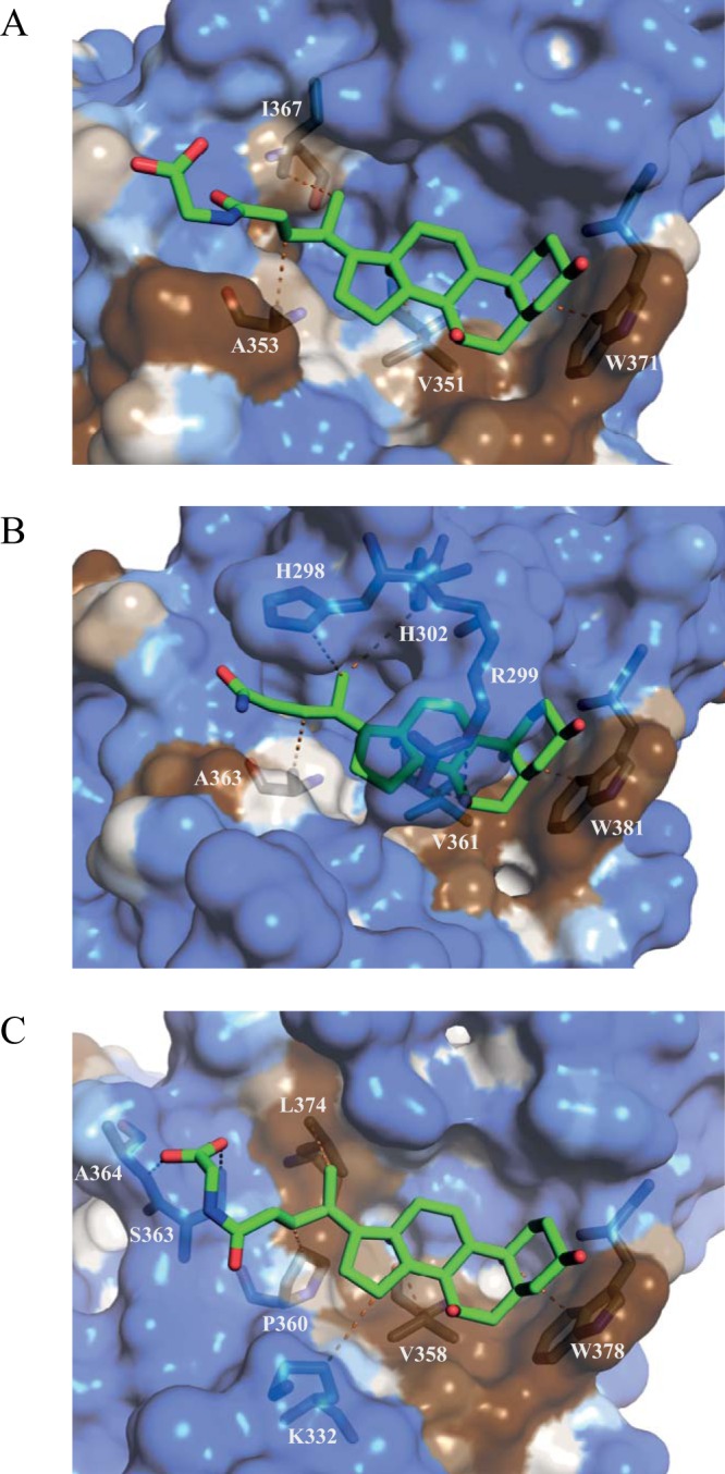
FIG 8.

Hydrophobicity at the GII bile acid binding pocket. Hydrophobic surface representations of each P domain, GII.1 (A) GII.10 (B), and GII.19 (C), in complex with bile acid (GCDCA) (green sticks) indicate that bile acids rest on a partly hydrophobic surface (brown) in the binding pocket. Hydrophilic regions are shown in blue. Only residues that interact directly with bile acid are shown. Residues that are involved in the water-mediated interactions are not shown.
