Figure 3.

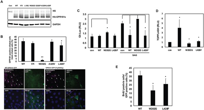
The functional assessment of GPR161 rare variants. (A) The protein expression pattern of GPR161 rare SNVs in 293 T cells. The cells were transfected with WT and six variants GPR161, and western blot analysis was performed with anti-HA and GAPDH antibodies. ns indicates a nonspecific band. (B) The ciliary localization of GPR161 rare SNVs in NIH3T3 cells. Upper panel shows the ratio of GPR161 positive cilia. GPR161 positive primary cilia were counted up to 200 cells from three independent experiments. Lower panel shows the ciliary localization with WT and p.Trp202Gly GPR161 (**P < 0.005, *P < 0.05). The arrows in the acetylated α-tubulin panels indicate the total ciliated cells, and the arrows in the merge panels indicate the GPR161 positive cilia. (C) The effect of GPR161 rare SNVs on Gli-responsive luciferase activity in C3H10T1/2 cells. The cells were transfected with Gli-luc and SV40-Renilla along with GPR161 (WT and SNVs) expression vectors and were treated with/without SAG for 24 h prior to lysis. Relative Luciferase Unit (RLU) indicates the relative Gli-luciferase activity over renilla luciferase (*P < 0.05). (D) The effect of GPR161 rare SNVs on the TOPFLASH luciferase activity in 293 T cells. The cells were transfected with TOPFLASH and pTK-Renilla along with GPR161 (WT and SNVs) expression vectors. RLU indicates the relative TOPFLASH luciferase activity over renilla luciferase (*P < 0.05). (E) The effect of GPR161 rare SNVs on the cell proliferation in Gpr161 KO MEF cells. The cells were transfected with GPR161 (WT and SNVs) expression vectors and were treated with BrdU for 12 h before fixation. The BrdU positive cells were counted and divided by the GFP positive cells (*P < 0.05).
