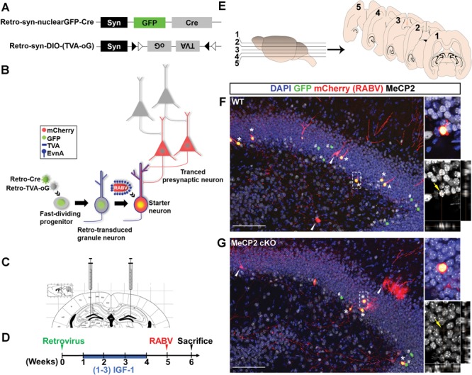
Figure 1.

Modified RABV-monosynaptic retrograde tracing of adult new DG neurons with targeted MeCP2 gene deleted. (A) Schematic illustration of dual-retroviral vectors for targeting Cre and Cre-dependent TVA (receptor for EnvA) and oG (rabies packaging protein) specifically to adult new neurons in the DG. (B) Schematic illustration of pseudotyped RABV-mediated monosynaptic retrograde tracing to map the presynaptic inputs to newborn neurons in the DG. Retrovirus infects only proliferating NSCs and progenitor cells in the adult DG and synapsin (syn) promoter ensures the transgenes are only expressed when NSCs differentiate into neurons. Retro-syn-nulcearGFP-Cre knocked out the MeCP2 gene in NSCs of cKO mice and labeled these cells with GFP. Retro-syn-DIO-(TVA-oG) expressed TVA and oG in Cre-dependent manner. TVA is the EnvA receptor, and EnvA is the envelope protein used for pseudotyping RABV. oG is required for RABV packaging and trans-synaptic spread. When pseudotyped RABV expressing mCh is injected, they can only infect the TVA+ cells. In these cells, the pseudotyped RABV replicated and trans-complementation with oG resulting in the production of mCh+ RABV that spread trans-synaptically to presynaptic (traced) neurons. Since presynaptic neurons do not have oG, no RABV production or further trans-synaptic transfer will occur. Therefore, starter cells are marked with both GFP and mCh while presynaptic neurons express only mCh. (C) Schematic illustration of virus injection into the DG. (D) Timeline of the RABV-mediated monosynaptic retrograde tracing experiment in vivo. (E) Schematic illustration of horizontal brain sections used for quantitative analysis. (F and G) Representative confocal images of a brain section containing the hippocampal region. Asterisks indicate the starter neurons (nuclear GFP+, mCh+). White arrows indicate traced presynaptic neurons (mCh+). Pounds indicate neurons that were infected by retrovirus but not RABV (GFP+). Immunohistological analysis shows that the starter neurons in DG of a WT mouse were positive for MeCP2 protein (F, yellow arrow), but the ‘starter neurons’ in a MeCP2 cKO mouse had no detectable MeCP2 signal (G, yellow arrows). Scale bar, 100 μm.
