Figure 2.

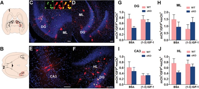
MeCP2 deletion in new DG neurons does not have significant effect on local hippocampal connection. (A and B) Schematic illustration of horizontal brain sections containing the hippocampus. (C–F) Representative confocal images of the red outlined area in (A) and (B) showing the connection between MeCP2-deficient DG new neurons with presynaptic neurons in DG, ML, CA3 and HL. Scale bar, 100 μm. (G–J) The ratio of traced cells (mCh+) to starter cells (GFP+ mCh+) in DG, ML, CA3 and HL were compared among experimental conditions: WT, n = 18; WT+ (1–3) IGF-1, n = 12; MeCP2 cKO, n = 14; MeCP2 cKO+ (1–3) IGF-1, n = 15. Two-way ANOVA with Tukey’s post hoc analysis for multiple comparisons; data were presented as mean ± SEM. There was no any significant difference in groups.
