Figure 3.

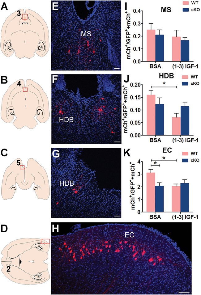
MeCP2-deleted new DG neurons have reduced long-range connectivity with the EC. (A–D) Schematic illustration of horizontal brain section through MS, HDB and EC. (E–H) Representative confocal images of the red outlined area in (A–D) showing the connection between MeCP2-deficient DG new neurons with presynaptic neurons in MS (I) and HDB (J) EC (K). Scale bar, 100 μm. (I–K) The ratio of traced cells (mCh+) to starter cells (GFP+ mCh+) in MS, HDB and EC were compared among conditions: MeCP2 deficiency in adult-born DG granule neurons reduced the ratio in EC (K, F (1, 55) = 2.299, P = 0.0467). (1–3) IGF-1 induced an obvious damage in normal newborn DG granule neurons in HDB (J, F (1, 55) = 5.958, P = 0.0149) and EC (K, F (1, 55) = 2.125, P = 0.0384). Two-way ANOVA with Tukey’s post hoc analysis for multiple comparisons; the numbers of animals used for quantitative analysis were WT, n = 18; WT+ (1–3) IGF-1, n = 12; MeCP2 cKO, n = 14; MeCP2 cKO+ (1–3) IGF-1, n = 15. Data were presented as mean ± SEM. *P < 0.05.
