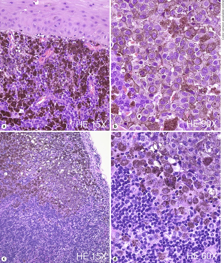
Fig. 3.
Histopathology findings. a Microscopic examination of the excised conjunctiva reveals a subepithelial proliferation of hexagonal-shape melanocytes with high nuclear/cytoplasmic ratio and very large nuclei. b These cells present a very irregular chromatin and frequent mitotic figures are noted. c, d Sentinel lymph node biopsy shows pigmented melanocytic cells with very atypical features consistent with epithelioid melanocytes and similar to the cells found in the conjunctival specimen.

