Fig. 1.
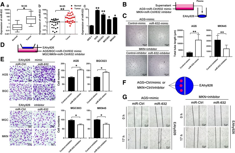
miR-632 is highly expressed in GC and improved GC cell angio-tube formation and endothelial cell recruitment. a The expression of miR-632 in GC tissues (a, n = 35) and serum (b, n = 25) from both the normal population and GC patients. (c) The expression of miR-632 was significantly higher in GC cells. b Schematic diagram showing the miR-632-mediated tube formation assay in GC cells. c Tube formation was accelerated by miR-632-mimic in AGS cells (upper left panels). miR-632-inhibitor decreased tube formation compared with the control in MKN45 cells (lower left panels). The histograms present the total tube length (mean ± SD) from three random fields at high magnification (right panels). d Schematic diagram showing the miR-632-mediated co-culture system using for endothelial cell Transwell assays in GC cells. e miR-632 mimic increased endothelial cell recruitment in AGS and BGC823 cells (upper left panels). The histograms present the cell numbers (mean ± SD) from three random fields at high magnification (upper right panels). miR-632-inhibitor suppressed endothelial cell recruitment in MGC803 and MKN45 cells (lower left panels). The histograms present the cell numbers (mean ± SD) from three random fields at high magnification (lower right panels). f Schematic diagram showing miR-632-mediated endothelial cell recruitment in wound healing assay of GC cells. g Wound healing was accelerated by miR-632-mimic in AGS cells (left panels). miR-632-inhibitor decelerated healing after scratching compared with the control in MKN45 cells (right panels). The experiments were performed at least three times independently. * P < 0.05; ** P < 0.01

