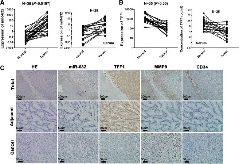
Fig. 2.
The expression of miR-632 was negatively associated with TFF1 in GC. a The expression of miR-632 in GC tissues compared with adjacent non-cancerous tissues (left panel, n = 35) and in GC serum compared with healthy serum (right panel, n = 25) measured via realtime PCR. b The expression of TFF1 in GC tissues compared with that in adjacent non-cancerous tissues measured by realtime PCR (left panel, n = 35). The concentration of TFF1 was detected via ELISA in GC serum compared with healthy serum (right panel, n = 25). c miR-632 expression was detected via in situ hybridization, and TFF1, MMP9 and CD34 expression was examined through immunohistochemiscal staining

