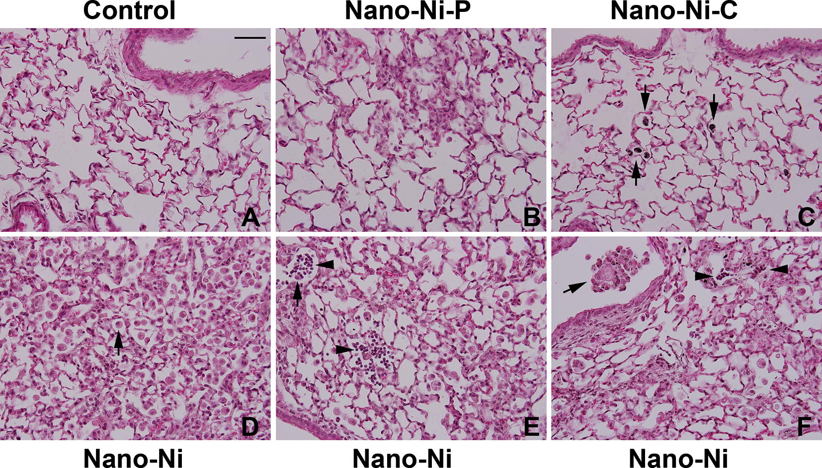
Fig. 7.

Histology in the lungs of mice 42 days after nickel nanoparticle exposure. Mice were instilled intratracheally with 50 µg per mouse of Nano-Ni, or with either partially passivated (Nano-Ni–P) or carbon-coated (Nano-Ni–C) nickel nanoparticles with same molar concentration of Ni as Nano-Ni. Control mice were instilled with physiological saline. Lung tissue sections collected from mice 42 days post-exposure were analyzed by H&E staining. A The normal structure of lung parenchyma in a control mouse. D–F Extensive chronic inflammation and fibrosis in the lungs of mice with Nano-Ni exposure. A large amount of enlarged and foamy macrophages in the alveolar space (arrow in D), alveolar septa, and lumen of bronchi and bronchioles (arrow in F), increased number of pulmonary intravascular macrophages (arrow in E), thickening of alveolar septa and subepithelial areas of bronchi and bronchioles, and lymphocytes infiltration (arrowheads in E, F) were observed. Chronic inflammation was also observed in Nano-Ni–P-exposed lungs, but at a much lesser degree (B). Only a mild chronic inflammation was observed in Nano-Ni–C-exposed lungs (C). Arrows in C: particle-phagocytized macrophages. Scale bar in A represents 50 µm for all panels
