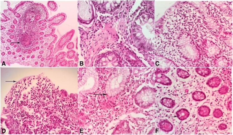
Fig. 1.
Histopathological lesions of Crohn’s disease, a Granulomatous ileitis: granuloma formation in the mucosa (arrow). b Mixed granulomatous enteritis is seen with Infiltration of lymphocytes and plasma cells in the mucosa. c Chronic lymphoplasmacytic enteritis: Infiltration of lymphocytes and plasma cells in the mucosa. d Epithelial patchy necrosis or ulceration of mucosal of ileum (arrow). e Pyloric gland metaplasia: compound acinar glands lined by antral type mucosa of stomach are observed in the lamina propria (arrow). f Paneth cell hyperplasia: These cells with numerous cytoplasmic granules are situated at the base of crypts of Liberkuhen glands (arrow) .H & E, ileum, human

