Figure 1.
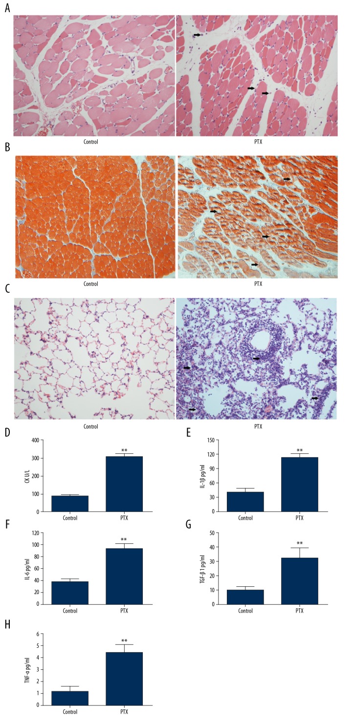
Histological analysis of the muscle and lung tissues. (A) Representative results of H&E staining of muscle tissue from mice treated with PBS or PTX. (B) Representative images of Masson’s trichrome staining of muscle tissue from mice in the PTX-induced inflammatory myopathies model. (C) Representative results of H&E staining of lung tissue from mice treated with PBS or PTX. (D) Serum levels of CK in mice treated with PBS or PTX. The level of IL-1β (E), IL-6 (F), TGF-β (G) and TNF-α (H) in mice treated with PBS or PTX. ** P<0.01.

