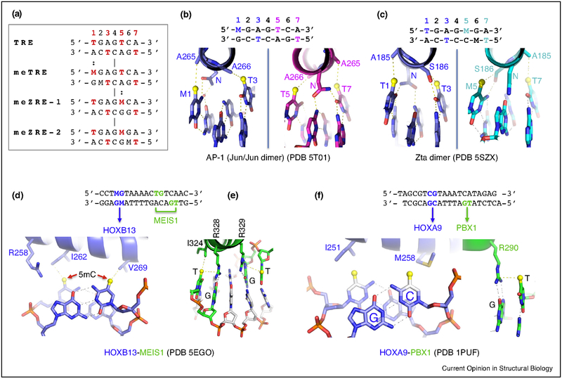
Figure 3.
Examples of methyl group recognition via van der Waals contacts. (a) Aligned DNA response elements with spatially equivalent methyl groups from T (5mU) or M (5mC). (b,c) Recognition of the four spatially constrained methyl groups of meTRE by Jun/Jun dimer (bZIP) or meZRE-2 by Zta dimer (bZIP). (d) Recognition of the two methyl groups of 5mCpG duplex by HOXB13 (homeodomain). (e) Homeodomain protein MEIS1 recognizes TpG via methyl–Arg–Gua triad. (f) HOXA9–PBX1 in complex with DNA. Both proteins could have preferential binding of methylated DNA. A methyl group (in yellow sphere) is modeled onto unmodified cytosine.

