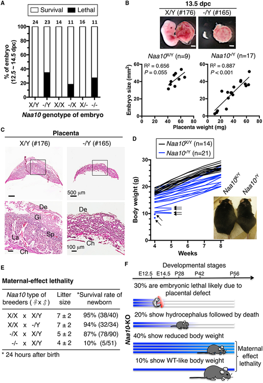
Figure 1. Phenotypes of Naa10-KO Mice.
(A) Naa10p deficiency causes partial embryonic lethality. Survival of Naa10-KO embryos at 12.5–14.5 days post-coitum (dpc) is shown. Naa10X/Y and Naa10−/Y are from Naa10−/X females crossed with WT or Naa10-KO males. Naa10X/X and Naa10−/X are from Naa10−/X females crossed with WT males. Naa10X/− and Naa10−/− are from Naa10−/X females crossed with Naa10-KO males. Embryos alive or dead were determined by heartbeat and embryo morphology. Embryo numbers are indicated above each bar.
(B) Positive correlation between embryo size and placenta weight in defective Naa10-KO embryos. Embryo size and placenta weight were measured for embryonic day 13.5 embryos of the WT (Naa10X/Y, n = 9) and mutant (Naa10−/Y, n = 17), derived from Naa10−/X females crossed with WT males. Representative images of embryos (176 and 165) and their placentae are shown. Embryo size is plotted versus placenta weight for each individual. The correlation (R2) and the p value for the Pearson analysis are shown.
(C) Naa10 KO results in placental defects. Representative images of H&E-stained WT (176) and Naa10-KO (165) placentae at 13.5 dpc are shown. Regions in black rectangles are enlarged at the bottom. De, decidual; Ch, chorionic plate; Gi, trophoblast giant cells; La; labyrinthine zone; Sp, spongioblast.
(D) Naa10-KO mice show growth retardation. The body weight of WT (Naa10X/Y, n = 14) and Naa10-KO (Naa10−/Y n = 21) mice was monitored for 4 weeks. Black arrows indicate mice that were found dead at the next time point. The image is representative of WT and Naa10-KO mice at the age of 25 weeks.
(E) Naa10-KO mice exhibit maternal effect lethality. Four types of breeding were carried out with parental strains of the indicated genotype. Pup survival was determined by heartbeat 24 hr after birth.
(F) Summary of phenotypes of Naa10-KO mice.
See also Figure S1 and Tables S1–S3.

