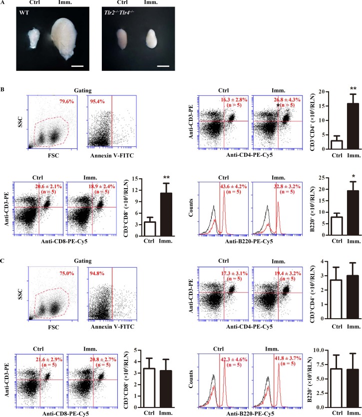
Fig. 4.

Lymphocytes in renal lymph node (RLN). A) RLN size. WT and Tlr2−/−Tlr4−/− mice were immunized. At 50 days after the first immunization (Imm.), RLNs were collected. Images are the representatives of the RLNs of five mice. Bar = 1 mm. B) Lymphocytes in the RLNs of WT mice. RLN cells were isolated from control (Ctrl) and immunized WT mice. Cells were labeled with annexin V-FITC and appropriate antibodies. Cell debris and apoptotic cells were gated out (upper left panels). Ratios and absolute numbers of CD3+CD4+ T cells (upper right panels), CD3+CD8+ T cells (lower left panels), and B cells (lower right panels) were determined using flow cytometry. C) Lymphocytes in RLNs of Tlr2−/−Tlr4−/− mice. RLN cells were isolated from control and immunized Tlr2−/−Tlr4−/− mice. Cells were analyzed as in B. Data are mean values ± SEM of five mice. Two RLNs per mouse were used in each flow cytometry analysis **P < 0.01.
