Fig. 5.

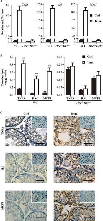
Expression of proinflammatory cytokines in the testis. A) Relative mRNA levels of cytokines. Total RNAs were extracted from testes of control and immunized WT and Tlr2−/−Tlr4−/− mice. Relative mRNA levels of major proinflammatory cytokines, including Tnfa, Il6, and Mcp1, were determined using real-time qRT-PCR normalized to β-actin. B) Protein levels of cytokines in the testis. Testes were lysed in PBS. The cytokine levels in the testis lysates of WT (left panel) and Tlr2−/−Tlr4−/− (right panel) mice were measured using ELISA. C) Cellular distribution of cytokines in the testis. Immunohistochemistry on the cryosections of testes from control (Ctrl; left panels) and immunized (Imm.; right panels) WT mice was performed using antibodies against TNFA, IL6, and MCP1. Insets in the upper right corner are the negative controls in which preimmune sera were used as primary antibodies. Arrows and arrowheads indicate Sertoli cells and interstitial cells, respectively. Images represent at least three independent experiments on three mice. Bar = 20 μm. Data represent mean values ± SEM of three experiments. **P < 0.01
