Fig. 6.

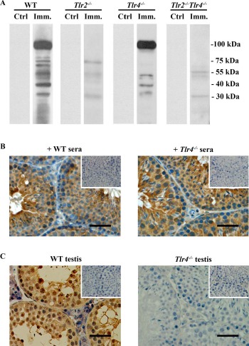
Autoantibody production. A) Autoantibodies in sera. Male germ cells were isolated from 10-wk-old WT mice. Western blot analysis on the germ cell lysates was performed using sera (1:500 dilution) of WT, Tlr2−/−, Tlr4−/−, and Tlr2−/−Tlr4−/− mice 50 days after the first immunization (Imm.) as the primary antibodies. The sera of mice that were injected with CFA alone served as controls (Ctrl). B) Distribution of autoantibodies. Testicular sections of 10-wk-old WT mice were immunostained with sera (1:100 dilution) of immunized WT (left panel) and Tlr4−/− (right panel) mice as the primary antibody. Insets in the upper right corners are negative controls in which the sera of control mice were used as the primary antibodies. C) Autoantibody deposition. Testicular sections of immunized WT (left panel) and Tlr4−/− (right panel) mice were directly stained with anti-mouse IgG antibodies. Signals in germ cells of WT mice indicate deposition of antibodies against germ cell antigens. Testes of the control mice served as the negative control (insets in upper right corners). Images are representatives of at least three mice. One testis in each mouse was used for immunostaining. Bar = 20 μm.
