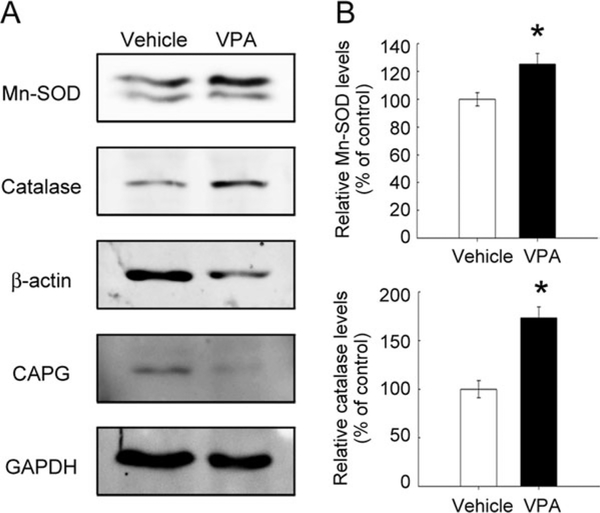
Fig. 3.
Expression of Mn-SOD and catalase in the lesion center of the spinal cord receiving VPA infusion. A: Proteins were extracted from the LC of the spinal cord at the indicated survival times after severe SCI, then subjected to Western blot analysis for the expression of β-actin and CAPG. B: Proteins were extracted from the LC of the injured spinal cords at day 7 post-SCI and then examined by Western blot analysis for Mn-SOD, catalase, β-actin, and CAPG protein levels. The relative intensity of Mn-SOD and catalase protein levels normalized to GAPDH was measured. Data are presented as mean ± SEM of percentage of the vehicle-treated control from three separate experiments. *P < 0.05 vs. vehicle.

