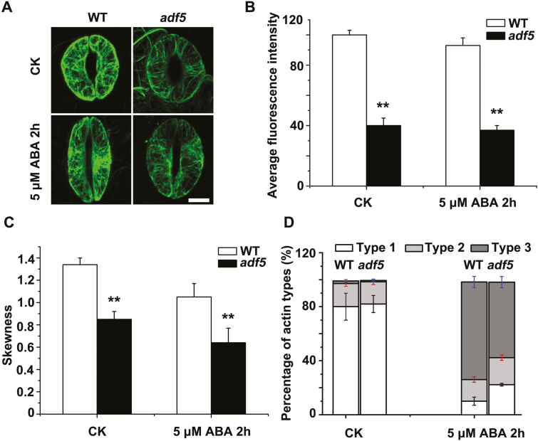
Fig. 4.
The loss of function of ADF5 delays actin reorganization during stomatal closure. (A) Confocal images of guard cells in rosette leaves from FABD2 (WT) and adf5×FABD2 transgenic plants after treatment with 5 μM ABA at 0 h (control, CK) and 2 h, showing GFP-labeled actin filaments. Scale bar=5 μm. (B) Quantification of the intensity of fluorescence of the GFP signal of WT and adf5 guard cells. (C) Quantification of bundling (skewness) of actin filaments in WT and adf5 guard cells. (D) Analysis of the type of actin organization in guard cells: type 1, radial array; type 2, random meshwork; type 3, longitudinal array. Data presented are the mean ±SE of three independent biological replicates. At least 60 stomata were analyzed for each time point. *P<0.05, **P<0.01 (Student’s t-test).

