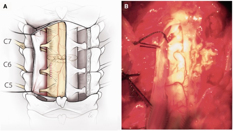
FIGURE 1.
Experimental model of neonatal brachial plexus injury. (A) Schematic showing the axial section of the spinal cord C5–C7 from the surgeon’s point of view where the dorsal rootlets and dorsal horns are superficial and the ventral roots and ventral horns are deep. The red dotted line represents the site of avulsion. (B) Intraoperative photograph of the surgical approach.

