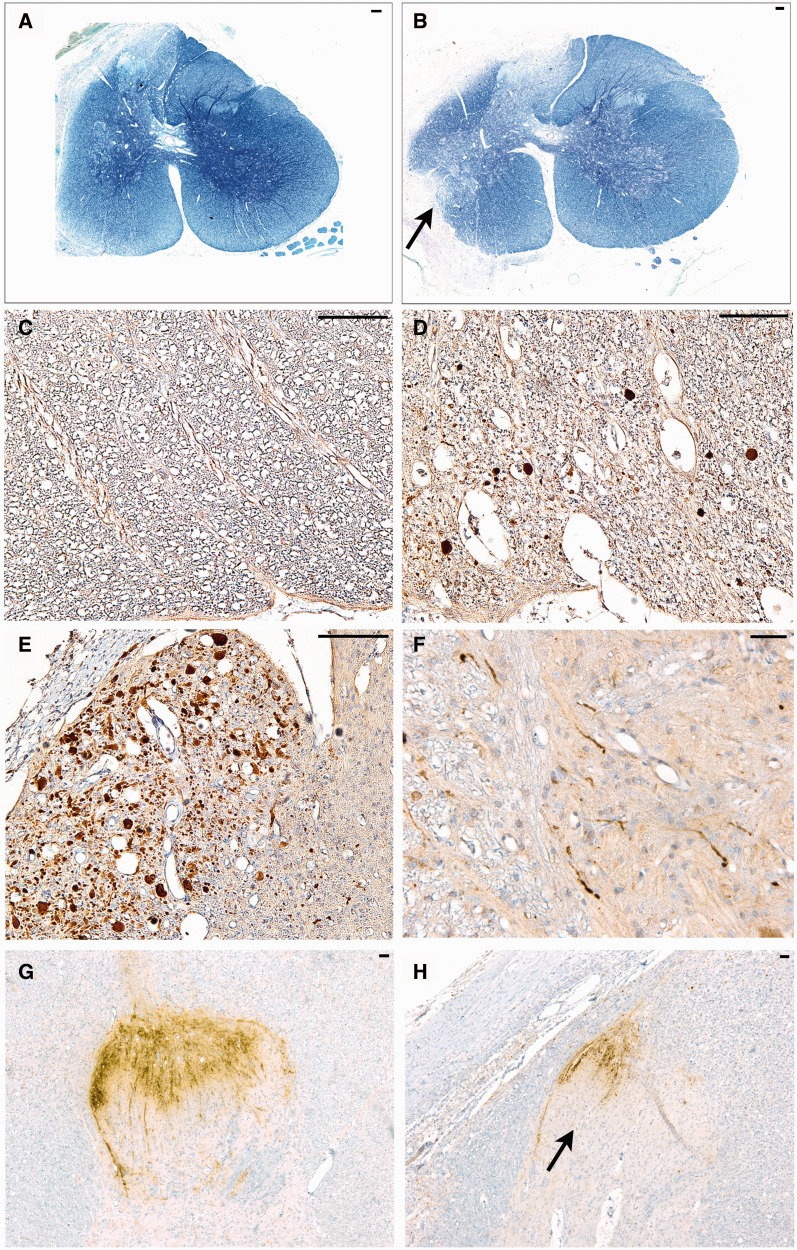
FIGURE 5.
Representative images of white matter injury and ongoing axonal degeneration at 6 weeks postinjury. (A) Whole Luxol fast blue (LFB)/Cresyl violet (CV)-stained cross-sectional C5 spinal cord following avulsion only. Note the pallor in myelin staining corresponding to the dorsal root entry zone (DREZ) and ventral root entry zone (VREZ) ipsilateral to avulsion with relative preservation of contralateral white matter integrity. (B) LFB/CV-stained cross-sectional C6 spinal cord following avulsion with myelotomy displaying marked pallor in myelin staining corresponding to extensive loss of white matter integrity in the DREZ and VREZ with relative preservation of contralateral white matter integrity. The arrow denotes the point of myelotomy. (C) An absence of amyloid precursor protein (βAPP)-positive axonal pathology in the C6 contralateral VREZ following avulsion with myelotomy. (D) C6 ipsilateral DREZ displaying swollen APP-positive profiles indicating ongoing axonal pathology following avulsion with myelotomy. (E) Extensive axonal pathology (βAPP-reactive) in the C7 VREZ ipsilateral to avulsion with myelotomy. (F) Swollen and varicose βAPP-reactive axons in the gray-white interface of the C5 ventral horn ipsilateral to avulsion with myelotomy. (G, H) Abundant calcitonin gene-related peptide (CGRP) immunoreactivity in sensory processes were observed on the contralateral side of the C6 level (G). This compared to CGRP immunoreactivity showing more marked ipsilateral loss of sensory processes in the C6 spinal cord level following avulsion with myelotomy (H). Scale bars: A, B, 100 µm; C–H, 50 µm.

