FIGURE 6.
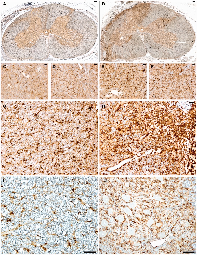
Whole spinal cord cross-section anti-Iba-1 stained sections demonstrate increased ipsilateral hemicord microglial activation following (A) avulsion only (C7) compared to the more extensive and widespread activation following (B) avulsion with myelotomy (C6). (C–F) High-magnification images display increased microglial clustering and reactivity in the C6 ipsilateral ventral horn following avulsion only (C), C6 contralateral ventral horn following avulsion only (D), C6 ipsilateral ventral horn following avulsion with myelotomy (E), and C6 contralateral ventral horn following avulsion with myelotomy (F). (G–J) Iba-1 immunoreactivity in C6 ipsilateral ventral root entry zone (VREZ) following avulsion only (G), C6 ipsilateral VREZ following avulsion with myelotomy (H), VREZ of naïve animal showing minimal Iba-1 immunoreactivity (I), and minimal Iba-1 immunoreactivity in the ventral horn of a naïve animal (J). Scale bars: A, B, J, 100 µm; C–I, 50 µm.

