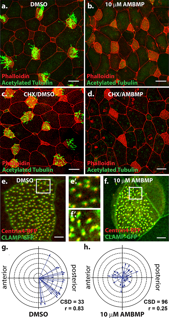
Figure 3: AMBMP blocks ciliogenesis and cilia polarization.
(a-b) Xenopus embryos stained with an antibody to acetylated tubulin (green) and phalloidin (red) treated with DMSO (a) or 10mM AMBMP (b) from stage 18–28 showing a complete loss of cilia in AMBMP treated embryos (b). (c-d) Similar experiment to a-b except embryos were pre-treated for 1 Hr with cycloheximide showing some cilia growth with DMSO (c) but not with AMBMP (d). (e-h) Representative images of Xenopus embryos injected with centrin4-RFP (red) and CLAMP-GFP (green) showing the loss of cilia polarity in embryos treated with 10mM AMBMP (f-f’) compared to DMSO (e-e’). (g-h) Cilia polarity is quantified with each arrow representing the data from a single cell such that the direction of the arrow represents the mean cilia orientation and the length of the arrow represents the variation. Scale bars = 25μm in a-d and 5 μm in e-f.

