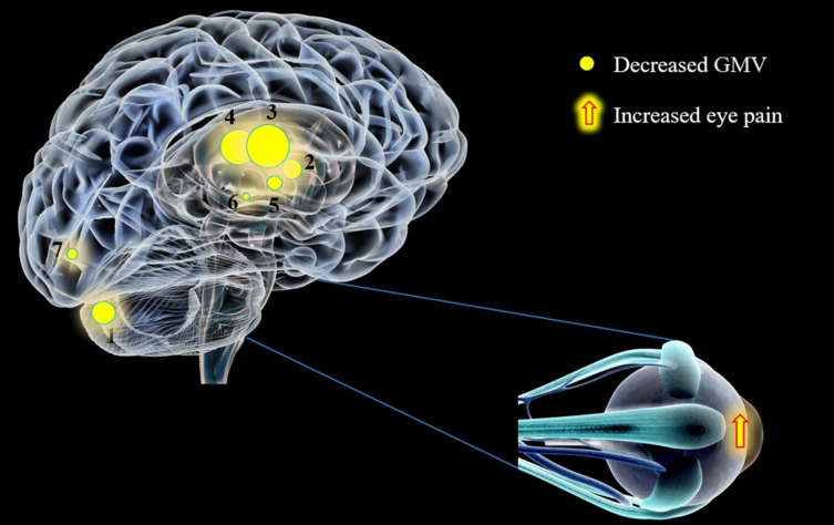
Figure 3.
The GMV values of altered brain regions in the EP group. Compared with the HCs, the GMV of the following regions were decreased to the following various extents: (1) cerebellum posterior lobe, L (t = −7.2225); (2) limbic lobe, L (t = −8.0419); (3) insula, R (t = −6.8695); (4) insula, L (t = −5.8375); (5) thalamus, L ( t = −9.7415); (6) caudate, L (t = −6.2255); and (7) cuneus, R (t = −5.1334) in EP patients. The sizes of the spots denote the degree of quantitative changes. L, left; R, right.

