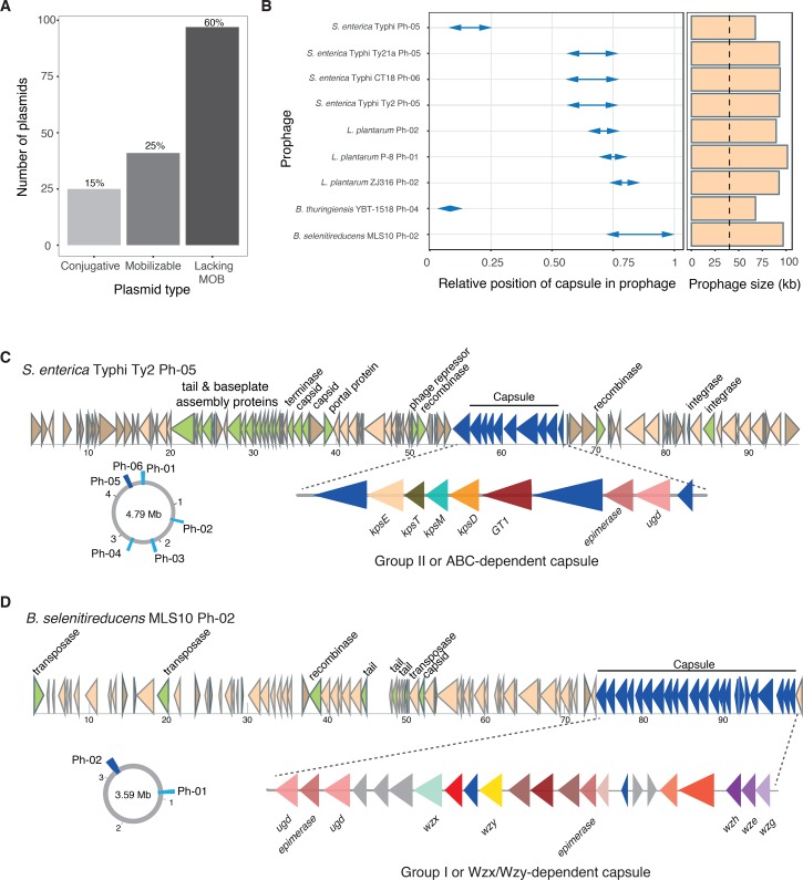
Fig 3. Capsule systems in MGEs.
A. Number of plasmids encoding capsule systems in function of the type of plasmid (classed in terms of mobility by conjugation). Plasmids lacking MOB may be mobilized by conjugation if they have a compatible oriT or mobilizable by other unknown means (e.g., natural transformation in competent species). B. Details of the nine capsules found in prophages. The arrows indicate the relative position and span of the capsule system in each prophage. Right panel indicates the size of each prophage. Dashed line indicated the average size of prophages in the database (40 kb). C and D. Details of the prophages and capsule systems from S. enterica and B. selenitireducens. Genetic schemes are drawn to scale (kb). In the drawing of the genetic locus of the prophage; genes associated to prophage biology are highlighted in green and capsule genes in dark blue. Circular diagrams represent the genomic localization of all the prophages in both species. The capsule-coding prophage is highlighted in dark blue. In the drawing of the locus of the capsule system, proteins in red-pink tones are associated to sugar modifications and may determine capsular serotype. Gene names are indicated below the arrows. GT1: glycosyl transferase.

