Figure 2.
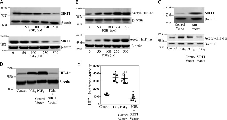
PGE2 increases HIF-1 activity by suppressing SIRT1 levels in human breast adipose stromal cells. A–E, human breast ASC cell line was used. A and B, the bottom panels represent primary human breast ASCs. A and B, cells were treated with the indicated concentrations of PGE2 for 24 h. Cells were then harvested, and lysates were subjected to Western blotting. C and D, cells were transfected with 2 μg of control vector or SIRT1 expression vector as indicated. C (top), cells were harvested, and Western blotting was performed for SIRT1 and β-actin to confirm overexpression. C (bottom) and D, cells were treated with vehicle or 500 nm PGE2 for 24 h. Cell lysates were prepared and subjected to Western blotting, and the blots were probed as indicated. E, cells were transfected as indicated with 0.9 μg of HRE-luciferase and 0.2 μg of psvβ-gal constructs. Cells labeled Control Vector also received 0.9 μg of expression vector; cells labeled SIRT1 Vector also received 0.9 μg of SIRT1 expression vector. 24 h after transfection, cells were treated with vehicle (control) or 500 nm PGE2 for 24 h. Cells were harvested, and luciferase activity was measured. Luciferase activity was normalized to β-gal activity. Means ± S.D. (error bars) are shown, n = 6. *, p < 0.001 versus PGE2-treated cells expressing control vector.

