Figure 4.
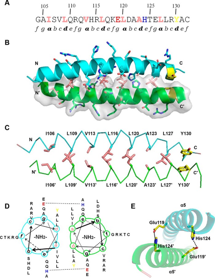
Dimerization interface of LZLM. A, primary amino acid sequence of α5 contains two partial and three complete repetitive heptad repeats (labeled as a–g). Hydrophobic residues at positions a and d are colored in pink and the C-terminal Tyr residue in yellow. Glu-119 and His-124 are showed in red and blue, respectively. B, dimerization interface of α5. One monomer is shown in cartoon representation and the other in surface rendering. Side chains of hydrophobic residues at positions a and d are shown as pink sticks. C, hydrophobic interactions and π-stacking at the dimerization interface. Side chains of hydrophobic residues at positions a and d are shown as pink sticks and π-stacking side chains in yellow. D, helical wheel diagram of α5. Glu-119 and His-124, which are involved in an interhelical salt bridge, are highlighted. E, detailed view of salt bridges between α5 and α5′. Glu-119 and His-124 of α5 form interhelical salt bridges with His-124′ and Glu-119′ of α5′, respectively.

